Dr. Black's Eye Associates of Southern Indiana
302 West 14th Street, Suite 100A
Jeffersonville, IN 47130
Phone: (812) 284-0660
Monday—Friday | 8 a.m.– 5 p.m.
Dr. Black's Eye Associates of Southern Indiana
302 West 14th Street, Suite 100B
Jeffersonville, IN 47130
Phone: (812) 284-1700
Monday—Friday | 8 a.m.– 5 p.m.
Retinal Detachment
What is a retinal detachment?
The retina is the light-sensitive layer of tissue that lines the inside of the eye and sends visual messages through the optic nerve to the brain. When the retina detaches, it is lifted or pulled from its normal position.
RETINAL DETACHMENT IS A MEDICAL EMERGENCY. If not promptly treated, retinal detachment can cause permanent vision loss.
If you have symptoms of a detached retina, it’s important to go to an eye doctor or the emergency room right away.
The symptoms of retinal detachment often come on quickly. If the retinal detachment isn’t treated right away, more of the retina can detach — which increases the risk of permanent vision loss or blindness.
Treatment for retinal detachment works well, especially if the detachment is caught early. In some cases, patients may need a second treatment or surgery if your retina detaches again.
What is a retinal tear?
In some cases there may be small areas of the retina that are torn. These areas, called retinal tears or retinal breaks, can lead to retinal detachment.
What are the symptoms of retinal detachment?
Symptoms include a sudden or gradual increase in either the number of floaters, which are little “cobwebs” or specks that float about in your field of vision, and/or light flashes in the eye. Another symptom is the appearance of a curtain over the field of vision.
Who is at risk for retinal detachment?
Anyone can have a retinal detachment, but some people are at higher risk. You are at higher risk if:
- You or a family member has had a retinal detachment before
- You’ve had a serious eye injury
- You’ve had eye surgery, like surgery to treat cataracts
Some other problems with your eyes may also put you at higher risk, including:
- Diabetic retinopathy (a condition in people with diabetes that affects blood vessels in the retina)
- Extreme nearsightedness (myopia), especially a severe type called degenerative myopia
- Posterior vitreous detachment (when the gel-like fluid in the center of the eye pulls away from the retina)
- Certain other eye diseases, including retinoschisis (when the retina separates into 2 layers) or lattice degeneration (thinning of the retina)
If you’re concerned about your risk for retinal detachment, talk with your eye doctor.
What causes retinal detachment?
There are many causes of retinal detachment, but the most common causes are aging or an eye injury.
There are 3 types of retinal detachment: rhegmatogenous, tractional, and exudative. Each type happens because of a different problem that causes your retina to move away from the back of your eye.
How can I prevent retinal detachment?
Since retinal detachment is often caused by aging, there’s often no way to prevent it. But you can lower your risk of retinal detachment from an eye injury by wearing safety goggles or other protective eye gear when doing risky activities, like playing sports.
If you experience any symptoms of retinal detachment, go to your eye doctor or the emergency room right away. Early treatment can help prevent permanent vision loss.
It’s also important to get comprehensive dilated eye exams regularly. A dilated eye exam can help your eye doctor find a small retinal tear or detachment early, before it starts to affect your vision.
How will my eye doctor check for retinal detachment?
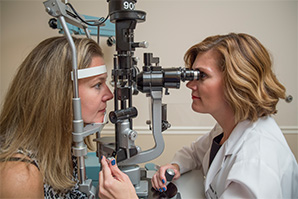
If you see any warning signs of a retinal detachment, your eye doctor can check your eyes with a dilated eye exam. Your doctor will give you some eye drops to dilate (widen) your pupil and then look at your retina at the back of your eye.
This exam is usually painless. The doctor may press on your eyelids to check for retinal tears, which may be uncomfortable for some people.
After a dilated eye exam, you may get an ultrasound or an optical coherence tomography (OCT) scan of your eye. Both of these tests are painless and can help your eye doctor see the exact position of your retina.


